
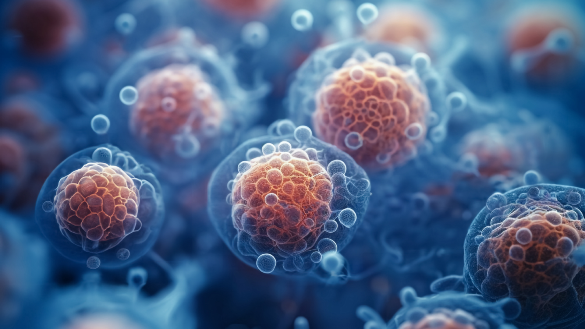
Cell Culture
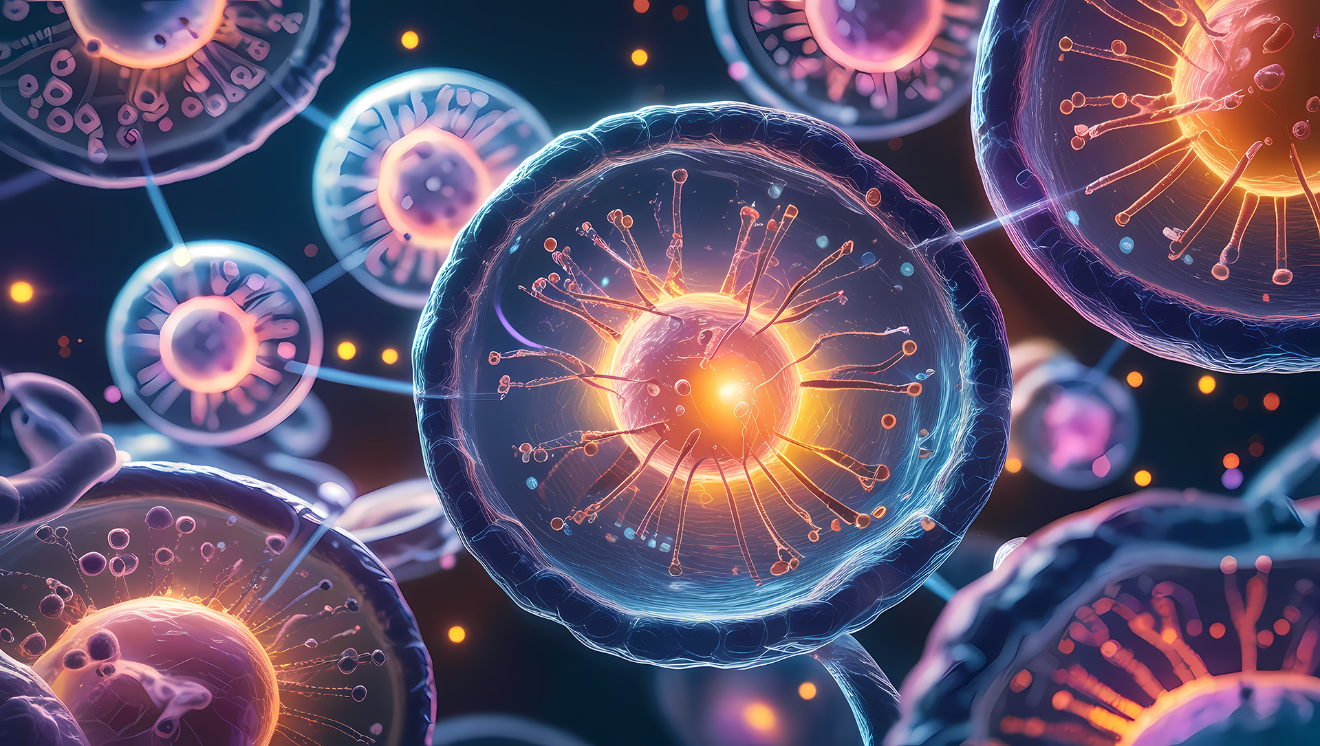
Multivesicular Body (MVB) Formation
Intraluminal Vesicle (ILV) Formation
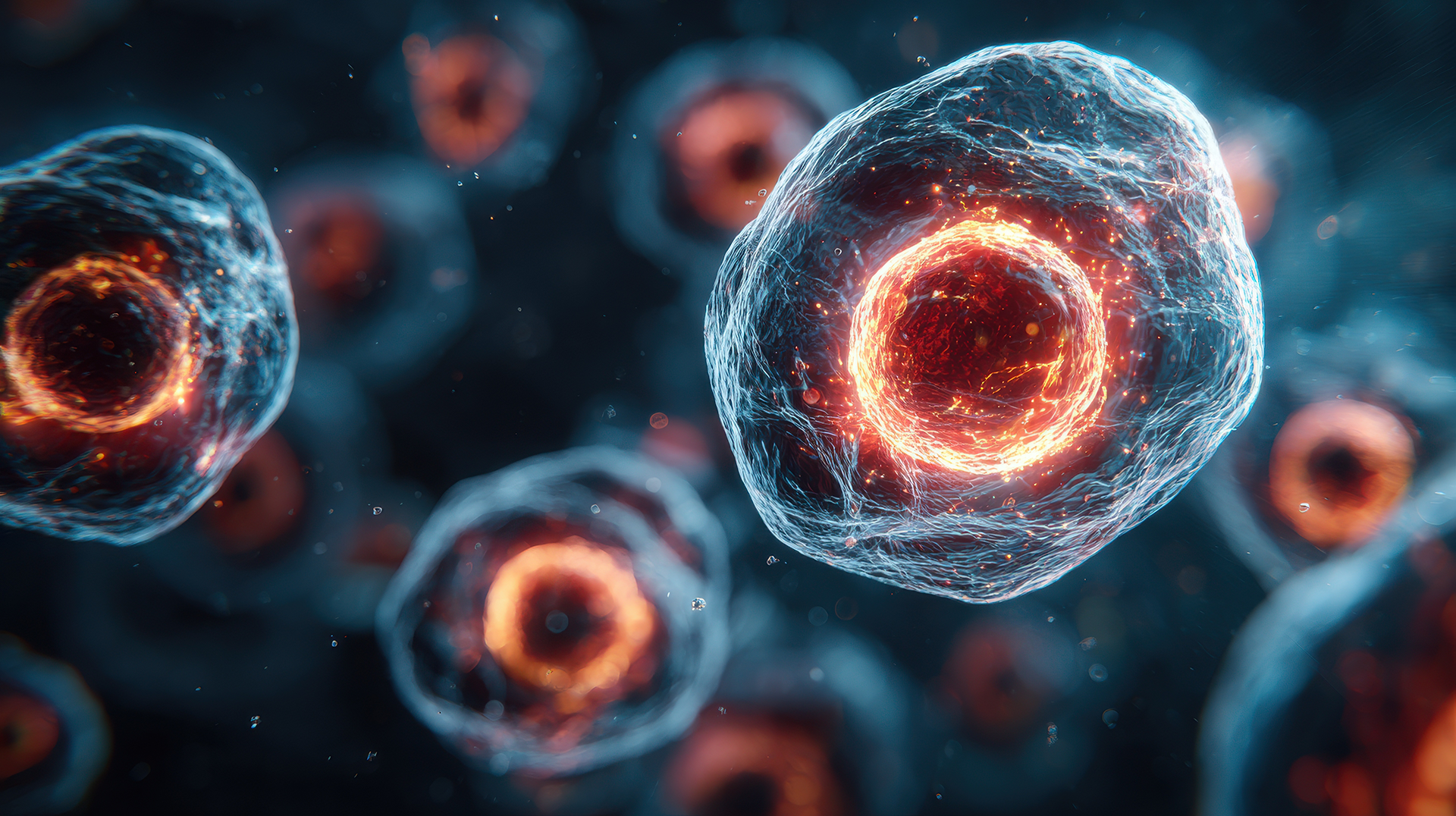
Secretion
Cell Culture
Most culture media used for the growth and propagation of cells in culture require the addition of fetal bovine serum (FBS) as a growth supplement.
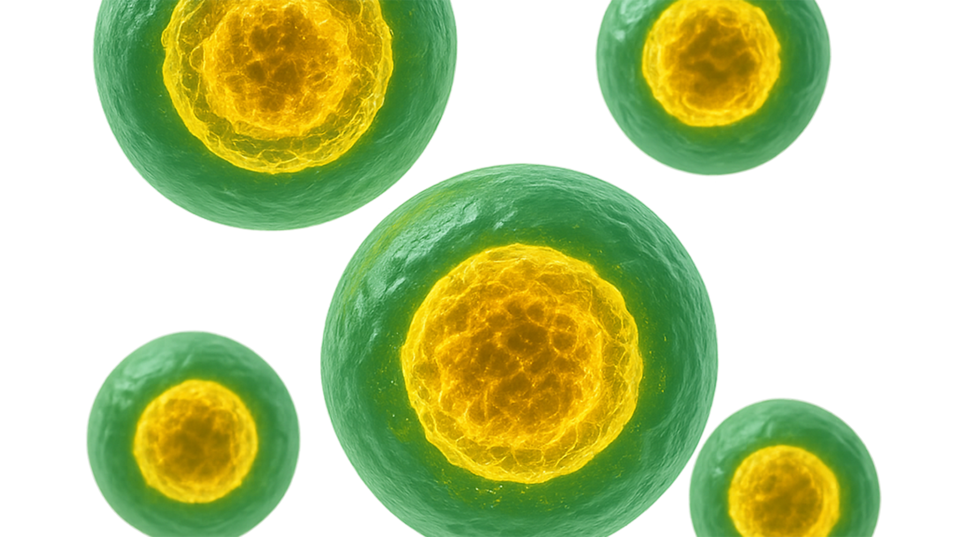
FBS is obtained from bovine (cow) serum, and therefore contains large quantities of cow exosome vesicles. These exosomes may interfere with some types of studies, or may lead to unreliable results when studying the exosomes shed from your cells of interest in normal culture conditions. Therefore, the use of exosome-depleted FBS is highly recommended for many types of studies.
Exosome depletion kits from Norgen in column and slurry format are also available.
Multivesicular Body (MVB) Formation.
Late endosomes can accumulate intraluminal vesicles (ILVs) by inward budding. MVBs sit at the crossroads of degradative (to lysosome) and secretory pathways, central to exosome biogenesis.
Implication for Isolation: Antibodies against CD63/CD81/CD9 may bias toward tetraspanin‑rich subsets. SEC and SmartSECTM preserve native membrane organisation; dUC offers flexibility to add cushions/gradients; exosome precipitation can increase yield, but co‑pulls soluble proteins, while SiC purification enables gentle capture with broad RNA recovery.
Bench tips: Keep cells healthy (EV‑depleted FBS, media‑only controls). If you must modulate output, GW4869 (nSMase2 inhibitor) often reduces ceramide‑dependent exosome release; calcium ionophores or bafilomycin A1 can boost release—use only for mechanism studies and report explicitly.
Secretion
Exosome release occurs when MVBs fuse with the plasma membrane. MVBs travel on microtubules then switch to actin for the final approach; Rab27a/b position and dock them via effectors like Munc13‑4 and the exocyst. Fusion is executed by R‑ and Q‑SNAREs (e.g., VAMP7/8/YKT6 with STX4/SNAP23), with Ca²⁺ acting as a key gatekeeper.
Release increases with calcium influx (e.g., ATP/P2X7), hypoxia/metabolic stress, and lysosome alkalinization, all of which can alter cargo. As mechanistic controls, use GW4869 (nSMase2) or knock down Rab27a/b, Munc13‑4, or YKT6—and always report any treatments and validation.
For conditioned media, use EV‑depleted FBS, typical 24–48 h windows, pre‑clear/filtration, and a media‑only control. Normalize as particles per 10⁶ cells per 24 h (NTA) and report positives (CD9/63/81, TSG101/ALIX) and negatives (Calnexin/GM130; albumin/ApoA1 for plasma). High‑release states can raise co‑secreted proteins—add an SEC/SmartSEC polish for proteomics. For low‑yield matrices, consider SiC pH‑dependent capture or polymer precipitation, then SEC/SmartSEC if purity matters.