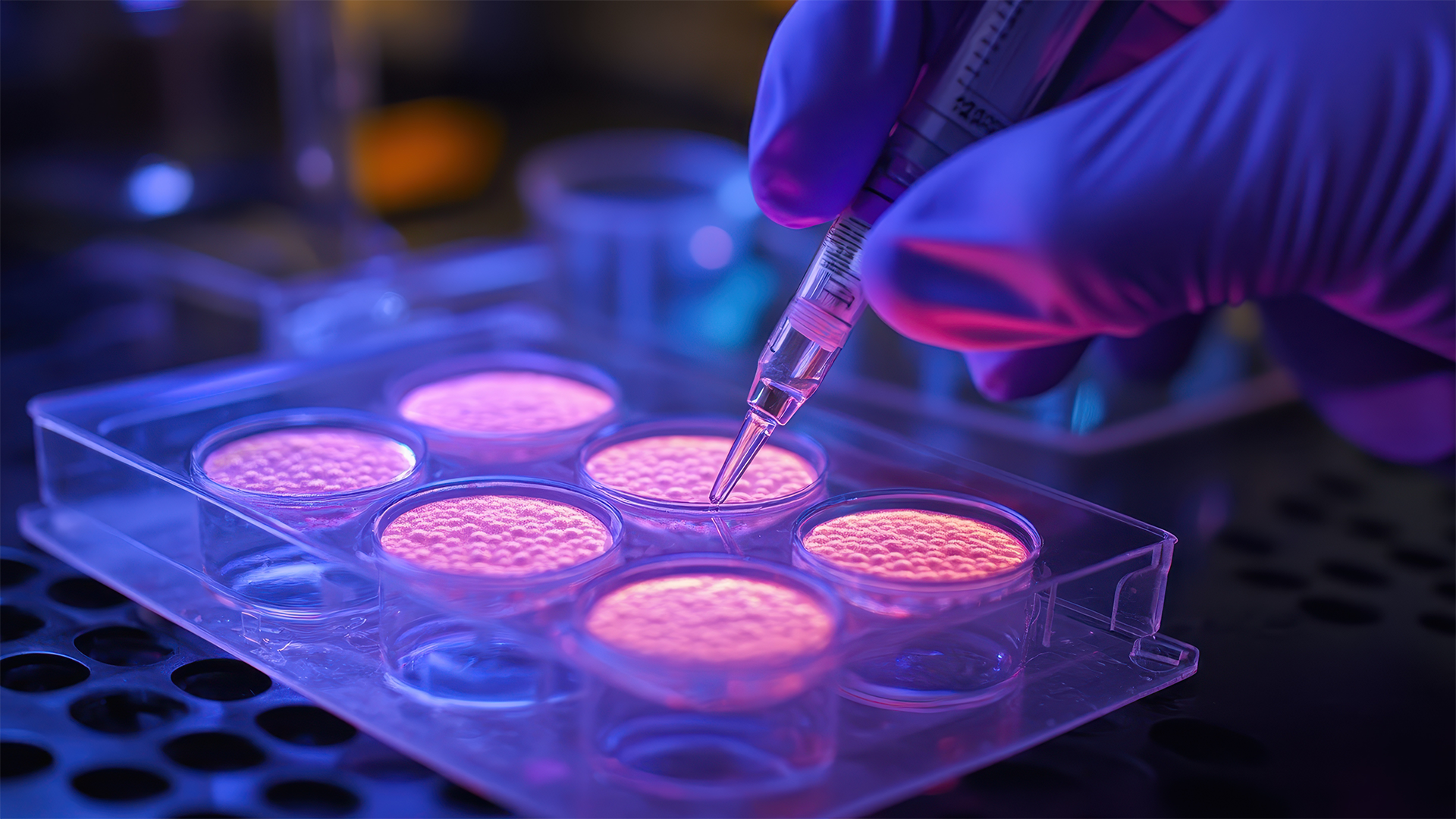

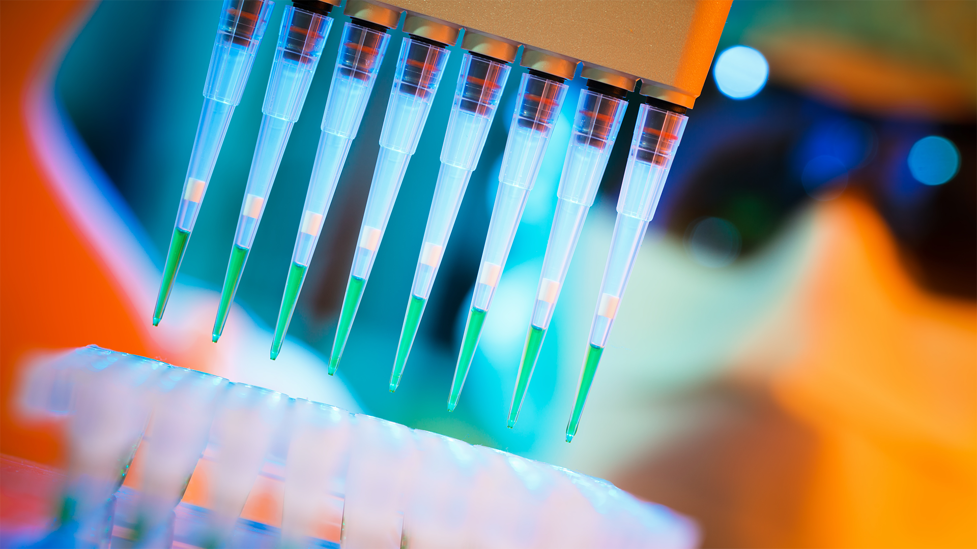
RNA Analysis & Profiling
Protein Mass Spectrometry
Lipidomics
NTA Particle Counting
Flow Cytometry

ELISA
RNA Analysis & Profiling
Advance your understanding of exosomal RNA with Norgen’s Exosome RNA Isolation Kit, engineered for maximal yield from plasma, serum, and cell culture supernatants, and Systems Bio’s SeraMir™ Exosome RNA Amplification System for downstream qPCR or NGS workflows.
Whether you’re troubleshooting low yield, selecting spike-in controls, or optimizing RNase-pretreatment, our RNA Analysis subpage guides you through best practices and normalization strategies to ensure reproducible profiling in miRNA- and lncRNA-focused studies.
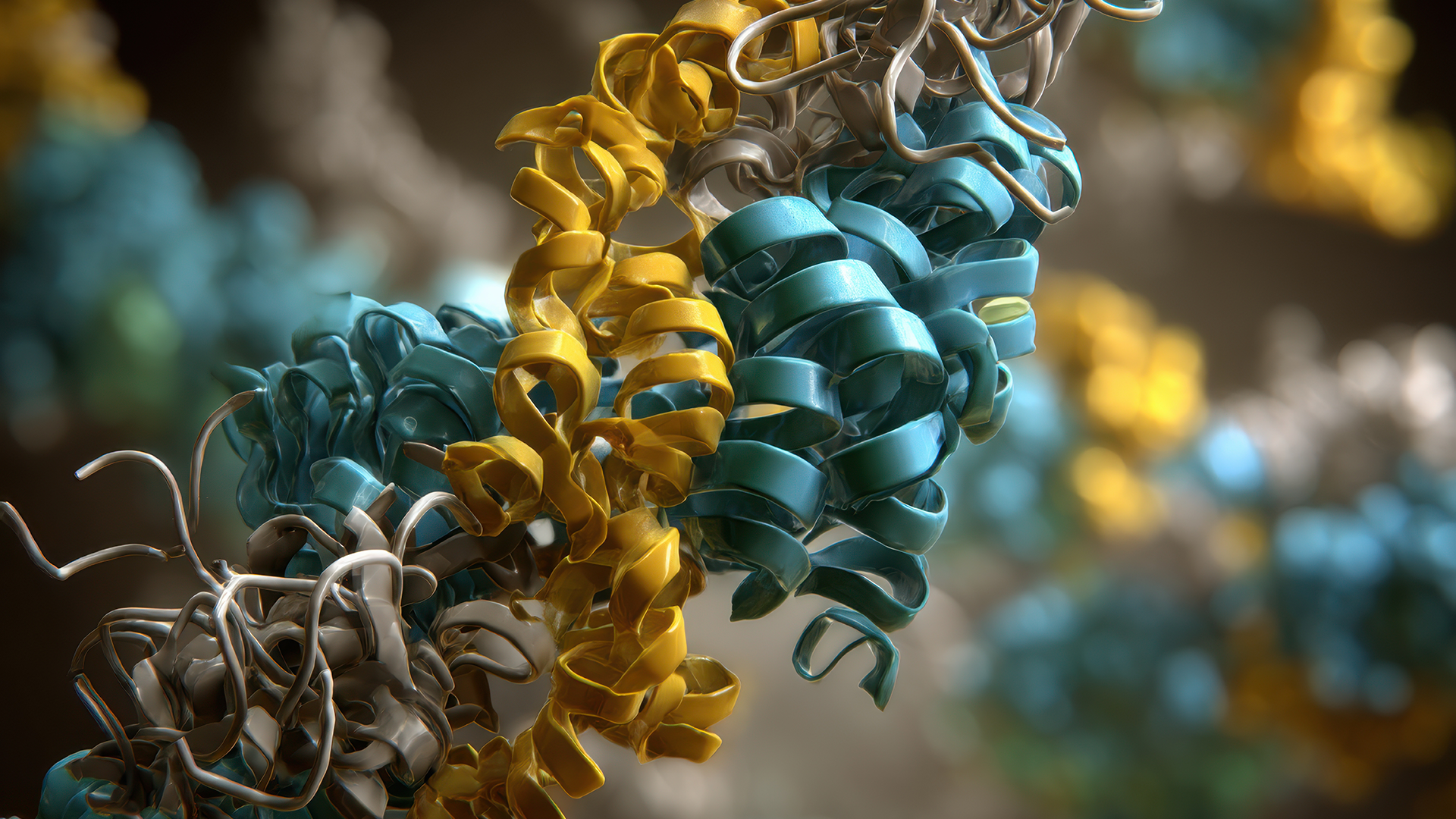
Protein Mass Spectrometry
Your complete workflow for protein-level insights.
The ExoMS™ line from System Biosciences makes extracellular vesicle protein analysis simple and reproducible. Whether you’re mapping the exosome from culture, serum, or plasma, ExoMS provides fast and clean capture for downstream discovery.
Why ExoMS?
-
Designed for compatibility with common EV isolation methods
-
Optimized for reproducibility across sample types
-
Streamlined workflows that save time and protect precious material
With ExoMS™, you can move seamlessly from vesicle prep to proteomic insight, turning your EV samples into meaningful biomarker data for discovery, validation, and translational research.
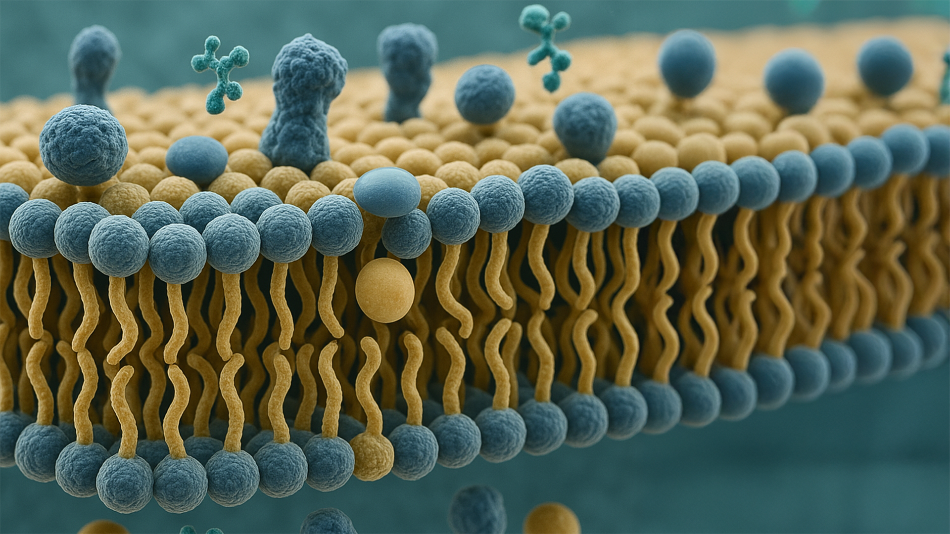
Lipidomics
Unlock the lipidome of your extracellular vesicles with our Lipidomics Services.
Exosomes carry the highest lipid-to-protein ratio among extracellular vesicles, making them a rich source of lipid biomarkers. Our LC-MS workflows sensitively profile sphingolipids, phospholipids, sterols, and more—giving you the resolution you need to truly decipher exosome biology.
You send us plasma, serum, or purified exosomes; we send back a clean dataset with putative IDs, m/z values, and fold-change analysis, ready to drop into your figures and downstream stats.
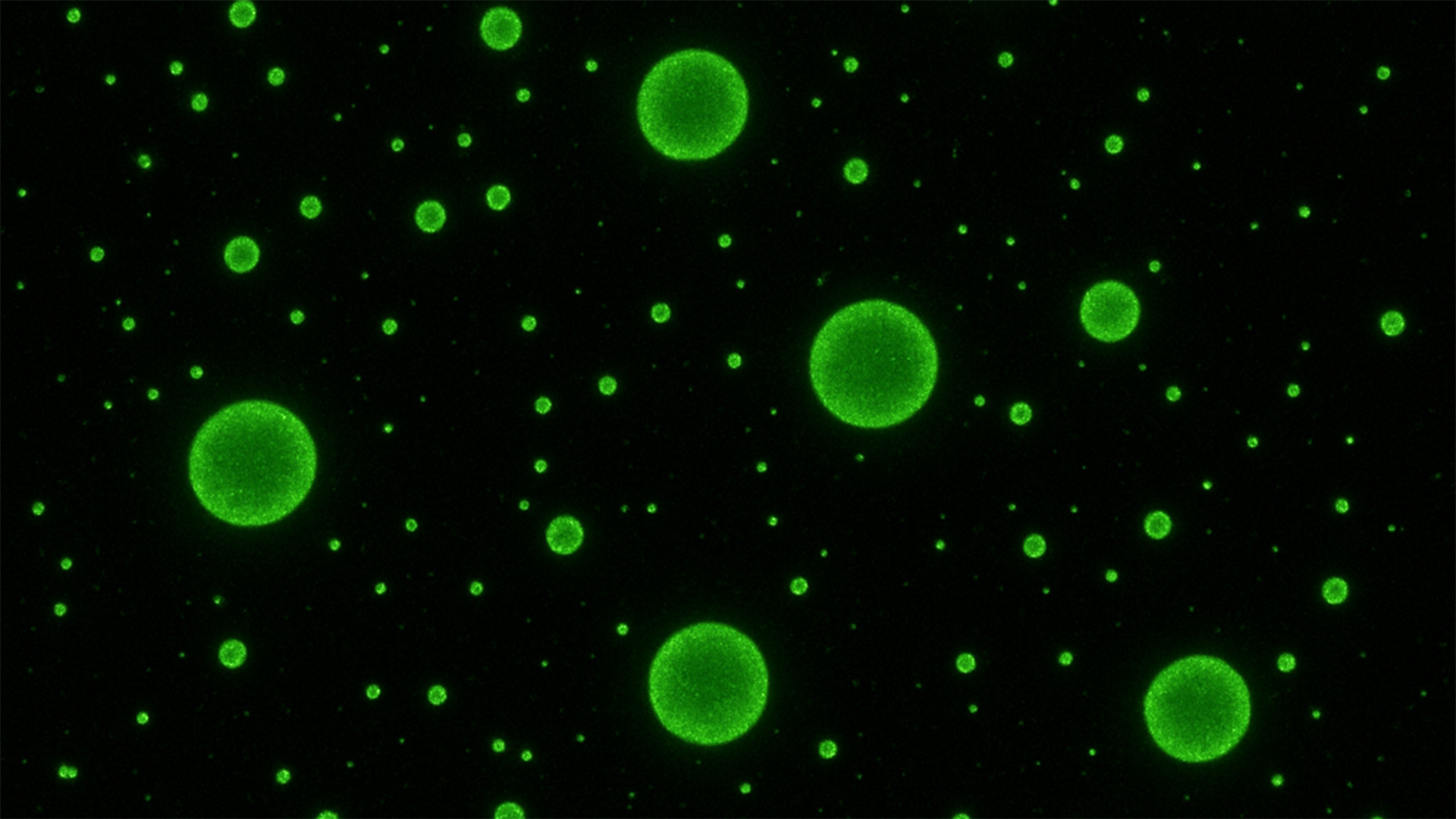
NTA Particle Counting
When particle counts matter for your grant reports and publications, you can’t afford background noise. Malvern’s NanoSight, paired with theExoGlow™-NTA Fluorescent Labelling Kit (the only commercially available EV-specific fluorescent labelling kit), gives you accurate size distribution and quantitation, without the uncertainty of non-vesicular contaminants.
-
See only EVs, not debris. Proprietary dye specifically labels extracellular vesicles, delivering high signal-to-noise you can trust.
-
Validated across workflows. Compatible with ExoQuick, ultracentrifugation, and SEC-based isolation.
-
Fast and reproducible. From isolated sample to analysis in just 30 minutes, optimized to keep your team moving.
This kit provides the confidence that your NTA readouts reflect vesicles, not artifacts.
Flow Cytometry
Flow cytometry was never designed for extracellular vesicles—until now. With SBI’s ExoFlow-ONE Gemstone dyes, you can finally unlock the full power of flow-based methods for EV biology.
-
Direct detection. Specifically labels internal EV components for clear, reliable signals.
-
Sharper data. Built-in size standards ensure every run is properly calibrated.
-
More insight per sample. Multiplex with spectrally separated dyes to ask more questions from the same prep.
-
Near single-vesicle resolution. Proprietary high-efficiency dyes reveal details once hidden in the noise.
The result? Flow cytometry that goes beyond bulk measurements—delivering sharper, cleaner, and more meaningful insights into EV biology.
ELISA
Looking for a sensitive, ready-to-run ELISA for exosomes or bacterial outer membrane vesicles (OMVs)? Discover the ExoELISA-ULTRA complete kits for quantitative detection of the key tetraspanins CD63, CD81, and CD9 in biofluids and cell culture supernatants—ideal for exosome characterization, biomarker validation, and QC of vesicle preps. Each kit is optimized end-to-end, from capture to colorimetric readout, so you get robust, reproducible data without building an assay from scratch.
Working with outer membrane vesicles? The ExoELISA-ULTRA GroEL kit enables specific detection of E. coli OMVs, while the BAMA kit is designed for Gram-negative bacterial OMV detection across strains. Upstream of ELISA, Norgen’s Bacterial EV Isolation Kit provides a fast, column-based workflow to purify bacterial EVs from culture supernatants, delivering clean OMVs ready for ExoELISA-ULTRA assays.
Together, these tools provide a unified ELISA toolkit for EV and OMV workflows—ideal for vaccine research, host–pathogen studies, and microbiome-related projects where you need quantitative, publication-ready data rather than another home-brew assay to optimize.